|
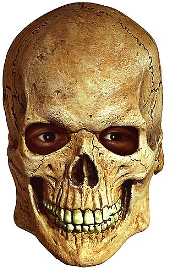
The human skull is
part of the skeleton, a bony structure which provides a strong cavity to
house our huge brain, a hinge for the jaw (mandible), to enable us to
chew and speak, and a support for the facial muscles and eyes, to be able to
see and communicate by expressions such as laughing - one of my favorites.
The human body is complex, consisting of various organs, muscles, ligaments, tissues and bones all with their own names and
functions. If you are considering a career in medical science, bionics
or robotics it is important that you have a
good understanding of the mechanics of the human body.
The
skull is not just another calcium casing, it is particularly interesting to map the development of Homo Sapiens
(modern humans). As we have developed from the high plains hominids,
such as Homo Habilis,
Homo Erectus, our cranial cavity grew to encompass the increase in brain
size, a clear indication of the need to cope with the increasing
complexity of life in social situations, the the need to adapt more
quickly that simple evolutionary advances could possible cope with. For
example, it would take millions (possibly billions) of years of trial
and error evolution before a Chimpanzee could build a jet fighter.
Whereas, modern humans have mastered that in a little less than 100
years using his brain.
The skull consists of 23
basic sections:-
-
Coronal
suture
-
Frontal
bone
-
Sphenoid
bone
-
Ethmoid
bone
-
Lacrimal
bone
-
Lacrimal
canal
-
Nasal
bone
-
Zygomatic
bone
-
Condyloid
process
-
Maxilla
-
Mental
foramen
-
Mandible,
body
-
Coronoid
process
-
External auditory
meatus
-
Mastoid
process
-
Mandibular
fossa
-
Zygomatic
process
-
Occipital
bone
-
Lambdoidal
suture
-
Temporal
bone
-
Squamosal suture
-
Parietal
bone
That said the adult human skull is said to consist of two different embryological origins: The neurocranium and the viscerocranium. The neurocranium (or braincase) is a protective vault surrounding the brain and brain stem. The viscerocranium (also splanchnocranium or facial skeleton) is formed by the bones supporting the face.
Except for the mandible, all of the bones of the skull are joined together by sutures, synarthrodial (immovable) joints formed by bony ossification, with Sharpey's fibres permitting some flexibility.
The Cranium
The skull or cranium is at the very top of the human body. To the untrained eye the skull may appear to be one large bone but
that is far too simplistic. The shape of the cranium has evolved
to protect human warriors from blows to the head.
Coronal Suture
The Coronal suture is a fibrous connective tissue joint which is rather dense which separates the parietal and frontal bones of the human skull.
At birth these bones of the skull do not meet.
Frontal bone
The frontal bone of the human skull bares a resemblance to a seashell and is made of two portions. These portions are the squama frontalis which is a vertical portion which corresponds with the forehead region and the Pars Orbitalis which is in an orbital or horizontal position and enters into the formation of the top of the nasal and orbital cavities.
Sphenoid bone
The Sphenoid bone (derived from the Greek: 'sphenoeides' meaning
wedge-like) is a singular bone in a shape resembling a butterfly which is located at the base of the human skull in front of both the temporal bone and basilar part of the occipital bone. This bone is one of seven which combine to form the orbit.
Ethmoid bone
The Ethmoid bone is located at the roof of the nose between the two orbits or eye sockets and is one of the bones which make up the orbit of the eye. This bone is responsible for separating the nasal
cavity from the brain and is cubical in shape and is of a cellular construction
to save weight.
Lacrimal bone
The smallest bone in the human face is the Lacrimal bone which is located at the front section of the medial wall of the eye socket.
Lacrimal canals
The Lacrimal Canals (also known as “Lacrimal canaliculi” or “Lacrimal ducts”) are the channels located in each eyelid which are small in size and
are seen as tiny orifices which are known as puncta lacrimalia.
Nasal bones
The Nasal bones in the human skull are two small sized bones which are oblong in shape
and vary considerably in size. These bones are beside one another in the upper and middle section of the face and form the “bridge” of the nose.
Body
The entirety of the skull is often referred to as the body of the skull.
Zygomatic bone
The cheekbone is known as the Zygomatic bone or malar bone. This bone is a paired bone of the skull and goes with the maxilla, temporal bone, frontal bone and the sphenoid one.
Condyloid process
This bone is part of the mandible and consists of two portions which are the condyle and the neck which is the constricted portion which is responsible for supporting it.
Maxilla
The maxilla is two bones which are fused along the palatal fissure to
form the upper jaw. It is similar to the lower jaw.
Mental foramen
The Mental foramen is the name given to one of two holes which are located on the anterior surface of the mandible
as a conduit for the mental nerves and blood vessels.
Mandible
The Mandible is the bone which forms the lower jaw and holds the lower teeth in place.
The lower inner surface of the neurocranium
Various sources provide different numbers for the count of constituent bones of the human
neuro - and viscerocranium. Different textbooks classify the bones of the human skull differently, e.g. they may
include bones that are usually considered neurocranial bones in their list of facial bones. Some textbooks count paired bones
(such as cheek bones) only once instead of twice. Some sources describe the
upper jaws left and right parts as two bones.
Some sources include the ossicles, three of which on each side are encased within the temporal bones, though these are also usually not considered part of the skull. Extra sutural bones may also variably be present, but they are not counted. For all of these reasons, it may not be
easy to reach agreement on an authoritative bone count for the neuro- and viscerocranium and the human skull. However, such discrepancies between various sources are only differences in how to classify and/or describe the anatomy of the human skull, and regardless of what classification/description is used, the basic anatomy remains the same. With that in mind, as one possible classification, the human skull could for example be said to consist of
22 bones instead of 23.
The skull also contains the sinus cavities, which are air-filled cavities lined with respiratory epithelium, which also lines the large airways. The exact functions of the sinuses are debatable; they contribute to lessening the weight of the skull with a minimal reduction in strength, they contribute to resonance of the voice, and assist in the warming and moistening of air drawn in through the nasal cavity.

The lower inner surface of the neurocranium- 11 weeks' fertilization age
Development of the skull
The skull is a complex structure; its bones are formed both by intramembranous and endochondral ossification. The skull roof, comprising the bones of the splanchnocranium (face) and the sides and roof of the neurocranium, is formed by intramembranous (or dermal) ossification, though the temporal bones are formed by endochondral ossification. The endocranium, the bones supporting the brain (the occipital, sphenoid, and ethmoid) are largely formed by endochondral ossification. Thus frontal and parietal bones are purely
membranous. The geometry of the cranial base and its fossas: anterior, middle and posterior changes rapidly, especially during the first trimester of pregnancy. The first trimester is crucial for development of skull
defects.
At birth, the human skull is made up of 44 separate bony elements. As growth occurs, many of these bony elements gradually fuse together into solid bone (for example, the frontal bone). The bones of the roof of the skull are initially separated by regions of dense connective tissue called "fontanels". There are six fontanels: one anterior (or frontal), one posterior (or occipital), two sphenoid (or anterolateral), and two mastoid (or posterolateral). At birth these regions are fibrous and moveable, necessary for birth and later growth. This growth can put a large amount of tension on the "obstetrical hinge", which is where the squamous and lateral parts of the occipital bone meet. A possible complication of this tension is rupture of the great cerebral vein of Galen. As growth and ossification progress, the connective tissue of the fontanelles is invaded and replaced by bone creating sutures. The five sutures are the two squamous, one coronal, one lambdoid, and one sagittal sutures. The posterior fontanel usually closes by eight weeks, but the anterior fontanel can remain open up to eighteen months. The anterior fontanel is located at the junction of the frontal and parietal bones; it is a "soft spot" on a baby's forehead. Careful observation will show that you can count a baby's heart rate by observing his or her pulse pulsing softly through the anterior fontanel.
Pathology
If the brain is bruised or injured it can be life-threatening. Normally the skull protects the brain from damage through its hard unyieldingness; the skull is one of the least deformable substances found in nature with it needing the force of about 1 ton to reduce the diameter of the skull by 1
cm. In some cases, however, of head injury, there can be raised intracranial pressure through mechanisms such as a subdural haematoma. In these cases the raised intracranial pressure can cause herniation of the brain out of the foramen magnum ("coning") because there is no space for the brain to expand; this can result in significant brain damage or death unless an urgent operation is performed to relieve the pressure. This is why patients with concussion must be watched extremely carefully.
Dating back to Neolithic times, a skull operation called trepanation was sometimes performed. This involved drilling holes in the cranium. Examination of skulls from this period reveals that the "patients" sometimes survived for many years afterward. It seems likely that trepanation was performed for ritualistic or religious reasons and not only as an attempted life-saving technique.
Craniometry and morphology of human skulls
Like the face of a living individual, a human skull and teeth can also tell, to a certain degree, the life history and origin of its owner. Forensic scientists and archaeologists use metric and nonmetric traits to estimate what the bearer of the skull looked like. When a significant amount of bones are found, such as at Spitalfields in the UK and Jōmon shell mounds in Japan, osteologists can use traits, such as the proportions of length, height and width, to know the relationships of the population of the study with other living or extinct populations.
The German physician Franz Joseph Gall in around 1800 formulated the theory of phrenology, which attempted to show that specific features of the skull are associated with certain personality traits or intellectual capabilities of its owner. This theory is now considered to be obsolete.
Sexual dimorphism
While in early life there is little difference between male and female skulls, in adulthood male skulls tend to be larger and more robust than female skulls, which are lighter and smaller, with a cranial capacity about 10 percent less than that of the
male. However, new studies show that women's skulls are thicker and thus men may be more susceptible to head injury than
women. The male body is larger than the female body, which accounts for the larger size of the male skull; proportionally, the male skull is about the same size as the female skull. Male skulls typically have more prominent supraorbital ridges, a more prominent glabella, and more prominent temporal lines. Female skulls generally have rounder orbits, and narrower jaws. Male skulls on average have larger, broader palates, squarer orbits, larger mastoid processes, larger sinuses, and larger occipital condyles than those of females. Male mandibles typically have squarer chins and thicker, rougher muscle attachments than female mandibles.
LINKS:
www.halloweencostumes.org/skeleton-skull-mask.html
www.skullsdirect.co.uk/
HUMANS:
OTHER
ANIMALS:
|
AMPHIBIANS |
Such
as frogs (class: Amphibia) |
|
ANNELIDS |
As
in Earthworms (phyla: Annelida) |
|
ANTHROPOLOGY |
Neanderthals,
Homo Erectus (Extinct) |
|
ARACHNIDS |
Spiders
(class: Arachnida) |
|
ARTHROPODS |
Crabs,
spiders, insects (phyla: Arthropoda) |
|
BIRDS
|
Such
as Eagles, Albatross
(class: Aves) |
|
CETACEANS
|
such
as Whales
& Dolphins
( order:Cetacea) |
|
CRUSTACEANS |
such
as crabs (subphyla: Crustacea) |
|
DINOSAURS
|
Tyranosaurus
Rex, Brontosaurus (Extinct) |
|
ECHINODERMS |
As
in Starfish (phyla: Echinodermata) |
|
FISH
|
Sharks,
Tuna (group: Pisces) |
|
HUMANS
- MAN |
Homo
Sapiens THE
BRAIN |
|
INSECTS |
Ants,
(subphyla: Uniramia class:
Insecta) |
|
LIFE
ON EARTH
|
Which
includes PLANTS
non- animal life |
|
MAMMALS
|
Warm
blooded animals (class: Mammalia) |
|
MARSUPIALS |
Such
as Kangaroos
(order: Marsupialia) |
|
MOLLUSKS |
Such
as octopus (phyla: Mollusca) |
|
PLANTS |
Trees
- |
|
PRIMATES |
Gorillas,
Chimpanzees
(order: Primates) |
|
REPTILES |
As
in Crocodiles,
Snakes (class: Reptilia) |
|
RODENTS |
such
as Rats, Mice (order: Rodentia) |
|
SIMPLE
LIFE FORMS
|
As
in Amoeba, plankton (phyla: protozoa) |
|
|

New
energy drinks for performers
..
Thirst for Life

330ml
Earth can - the World in Your Hands
|